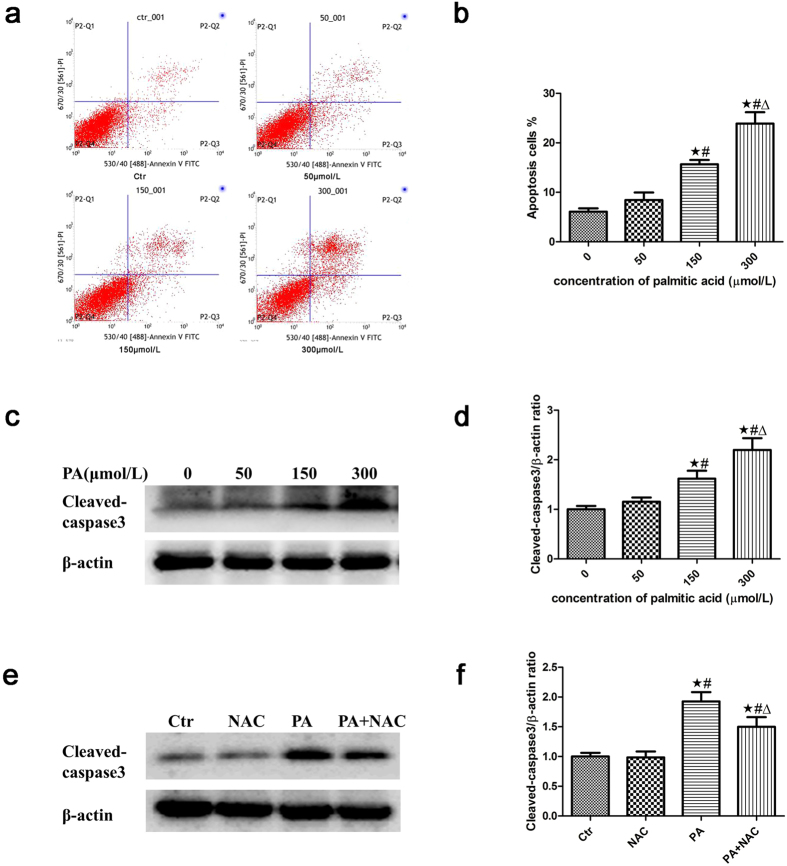
Figure 5. Palmitic acid induced apoptosis in podocytes.
Representative images of flow cytometry analysis of podocytes after treatment with different concentrations of palmitic acid for 24 hours. (a) Representative cytograms. (b) Percentage of apoptotic cells. ∗p < 0.05 vs. 0 μmol/L, #p < 0.05 vs. 50 μmol/L, Δp < 0.05 vs. 150 μmol/L. (c) Representative Western blot analyses of cleaved-caspase3 expression in podocytes treated with different concentrations of palmitic acid for 24 hours. (d) Densitometric analysis of cleaved-caspase3 expression in Figure c. ∗p < 0.05 vs. 0 μmol/L, #p < 0.05 vs. 50 μmol/L, Δp < 0.05 vs. 150 μmol/L. (e) Representative Western blot analyses of cleaved-caspase3 expression in podocytes treated with 150 μmol/L palmitic acid in the absence or presence of 150 μmol/L NAC for 24 hours. The cell lysates were analyzed by immunoblot using an antibody against cleaved-caspase3. (f) Densitometric analysis of cleaved-caspase3 expression in Figure e. The data are expressed as the mean ± SEM; n = 3. ∗p < 0.05 vs. control group, #p < 0.05 vs. NAC group, Δp < 0.05 vs. PA group. Ctr: Control group, podocytes were treated with 1% BSA. NAC: Podocytes were treated with 150 μmol/L N-acetyl-cysteine (NAC) for 24 hours. PA: Palmitic acid group, podocytes were treated with 150 μmol/L palmitic acid for 24 hours. PA + NAC: Podocytes were treated with 150 μmol/L palmitic acid for 24 hours after pretreatment with 150 μmol/L N-acetyl-cysteine (NAC) for 2 hours.

