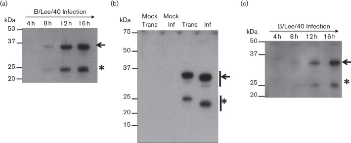
Fig. 1.
SDS–PAGE analysis of influenza B NS1 protein. (a). Time course infection of B/Lee/40 on HEK 293T cells. Cells were harvested at 4, 8, 12 and 16 h post infection and immunoblotted with αNS1. (b). HEK 293T cells were infected with B/Lee/40 virus (inf) or transfected with pCAGGS/NS1B-FLAG (Trans) and after 20 h the cell lysates were immunoblotted with αNS1. Mock Trans: Cells transfected with empty pCAGGS vector. Mock Inf: non-infected cells. (c). pCAGGS/NS1B-FLAG-transfected HEK 293T cell lysates were harvested at 4, 8, 12 and 16 h post transfection and immunoblotted using anti-FLAG. The full length (black arrow) and smaller (*) NS1 proteins are indicated.

