


Figure 7.
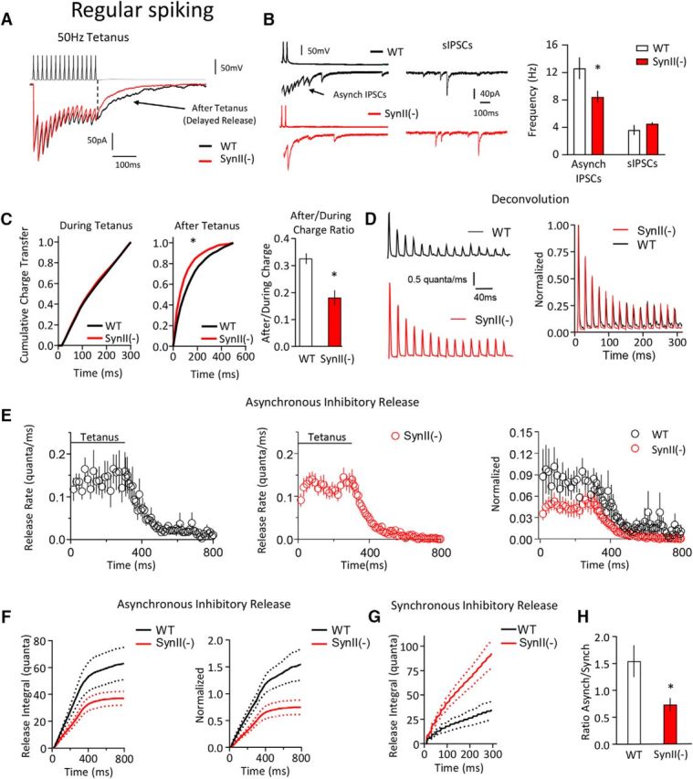
SynII deletion suppresses the asynchronous release component at regular-spiking (presumed CCK) interneurons. A, Representative traces illustrating that the after tetanus current is decreased at SynII(−) neurons. B, Asynchronous IPSC frequency is significantly (p < 0.05) reduced at SynII(−) neurons, whereas the sIPSC frequency is not affected. Left, Representative traces show asynchronous and spontaneous IPSCs. C, Cumulative probability plots show that SynII has no effect on the charge transfer during the tetanus (left), whereas the charge transfer after the tetanus is significantly reduced at SynII(−) neurons (middle, the distribution is shifted to the left, p < 0.05, Kolomogorov–Smirnov test). The After/During charge ratio is significantly decreased at SynII(−) neurons (right, p < 0.05). D, The synchronous release component at WT and SynII(−) neurons computed using the deconvolution. Right, Plot represents data normalized by the first response in a tetanus. E, The asynchronous release component computed using deconvolution. Right, Plot represents data normalized by the first response in a tetanus. F, The cumulative asynchronous release component is significantly decreased at SynII neurons (p < 0.05). Dotted lines indicate the confidence interval. G, The cumulative synchronous release component is significantly increased at SynII neurons (p < 0.05). Dotted lines indicate the confidence interval. H, The ratio between the cumulative asynchronous and synchronous release components is reduced at SynII(−) neurons (p < 0.05). Data collected from six pairs for each genotype. *p < 0.05.