Figure 2.

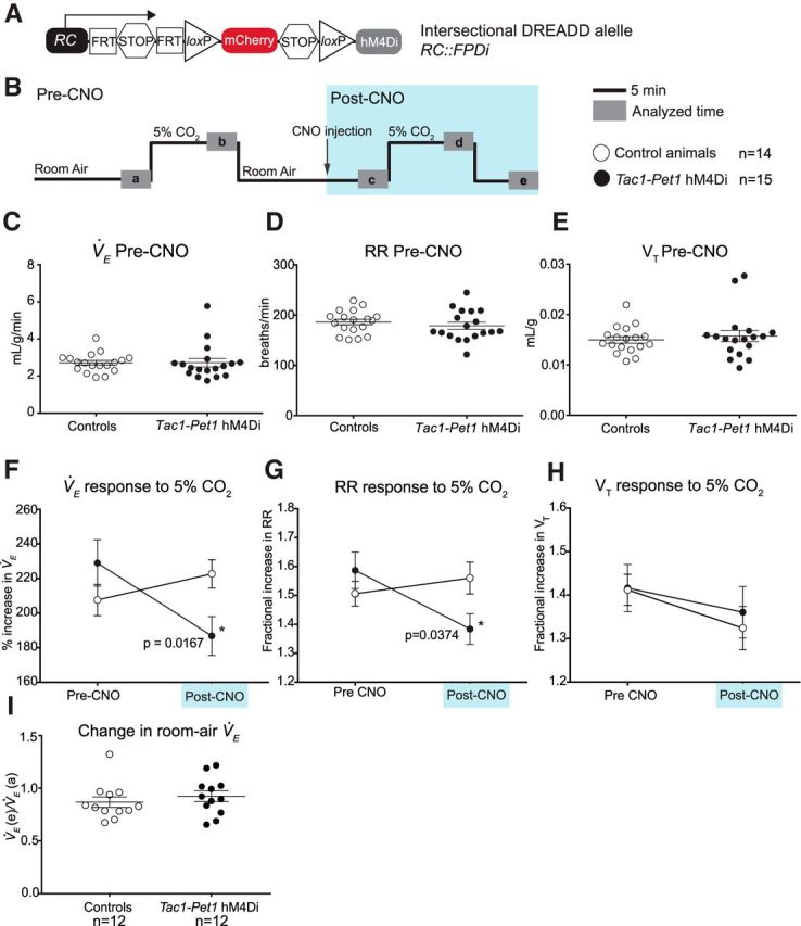
Acute silencing of Tac1-Pet1 neurons results in a decrease in ventilation at 5% CO2. A, Intersectional DREADD allele. B, Experimental design. After an initial period of acclimation, experimental adult animals and littermate controls are exposed to 5% CO2 and then returned to room air, briefly removed from the chamber for intraperitoneal injection of 10 mg/kg CNO, and placed back in room air. Animals were then exposed to 5% CO2 a second time, followed by another period of room air breathing in 12 animals from each group. Gray boxes (a–d) indicate time intervals used for data analysis. C–E, Baseline measurements of V̇E, respiratory rate (RR), and tidal volume (VT) in control (n = 14) and Tac1::IRES-cre, Pet1::Flpe, RC::FPDi (“Tac1-Pet1 hM4Di”) animals (n = 15) before CNO injection were not significantly different. Each circle represents one animal, error bars represent SEM. F, Average percentage increase in V̇E before CNO injection (pre-CNO, V̇Eb/V̇Ea × 100%) and minutes after CNO injection (post-CNO, V̇Ed/V̇Ec × 100%) in control and Tac1-Pet1 hM4Di animals. Error bars indicate SEM. G, H, Average fractional increase in respiratory rate (RR) or tidal volume (VT) to 5% CO2 before and after CNO injection. I, Change in room air ventilation after CNO administration compared with baseline (V̇Ee/V̇Ea) in control (n = 12) and Tac1-Pet1 hM4Di animals (n = 12). See Results for numerical data and statistical tests.
