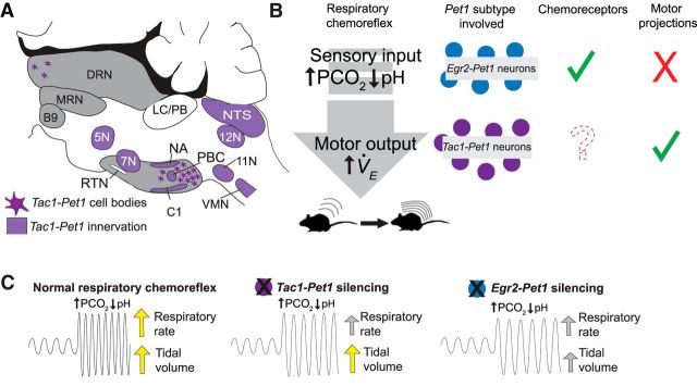
Figure 6.
Summary of Tac1-Pet1 innervation, cell body locations, and Pet1 neuron subtypes in the respiratory chemoreflex. A, Schematic illustrating regions receiving Tac1-Pet1 projections throughout the brainstem and spinal cord and locations of cell bodies within the raphe nuclei. B, Contrasting roles of Egr2-Pet1 and Tac1-Pet1 neurons in respiratory chemoreflex. Egr2-Pet1 neurons have been shown to respond directly to decreased pH, whereas direct recordings of Tac1-Pet1 neurons have not yet been performed. Both Tac1- and Egr2-Pet1 neuron subsets project to chemosensory regions, whereas only Tac1-Pet1 neurons project to motor nuclei within the brainstem. C, Effect of silencing Egr2-Pet1 and Tac1-Pet1 subsets on the respiratory rate and tidal volume components of the respiratory chemoreflex.

