
Figure 5.
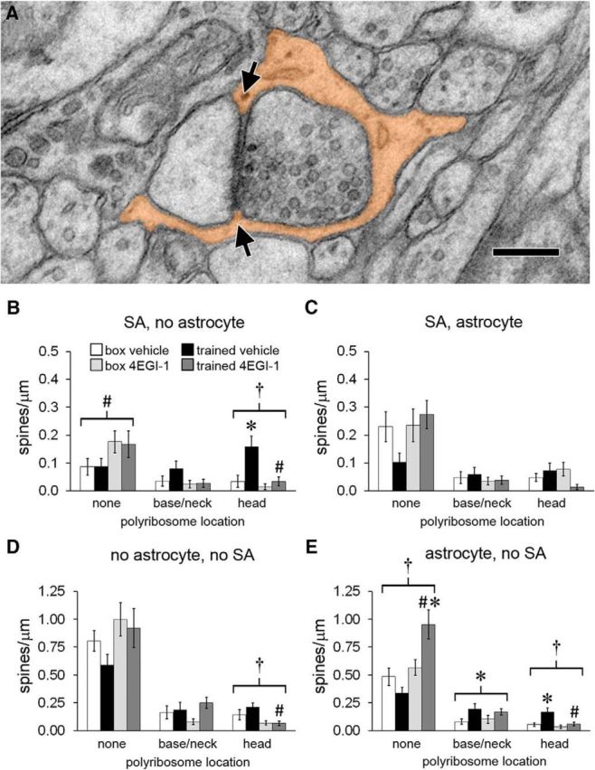
Astrocytes at synapses. A, Electron micrograph of a spine head with its synapse surrounded by an astrocytic process (orange) that makes contact with the synaptic cleft at both edges (arrows). Scale bar, 250 nm. B, For spines with a spine apparatus (SA) and no perisynaptic astrocyte, there was a main effect of drug on spines with no polyribosomes and an effect of training on spines with polyribosomes in the head that was blocked by the drug. C, There were no effects of training or drug on the frequency of spines with a spine apparatus and perisynaptic astrocyte. D, There were fewer spines with head polyribosomes but no spine apparatus or perisynaptic astrocyte in the trained-4EGI-1 group versus the trained-vehicle group. E, In spines with a perisynaptic astrocyte but no spine apparatus, there were more spines without polyribosomes in the trained-4EGI-1 group versus the box-4EGI-1 group and the trained-vehicle group. There was a main effect of training on spines with base or neck polyribosomes and an effect of training on spines with head polyribosomes that was blocked by the drug. Shown are effects at p < 0.05 for training (*), drug (#), and training × drug (†).