Figure 5.
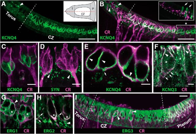
Immunoreactivities for KCNQ and ERG potassium channel subunits are observed in the turtle crista. A, B, Confocal image projection of a longitudinal section of turtle crista stained with antibodies against KCNQ4 subunit (green) and CR (magenta). White dashed line demarcates boundary between the torus and CZ. Arrowheads point to KCNQ4-positive afferent bouton terminals. Asterisk in B identifies group of CD afferents where CR staining is less intense or absent. A, Inset, Schematic of turtle hemicrista showing general plane of tissue section (red dashes) and zonal boundaries (black dashes) where T and P label the torus and planum, respectively. B, Inset, Confocal image projection of longitudinal section of the torus from another turtle crista stained with antibodies against KCNQ4 (green) and CR (magenta). CR stains bouton terminals and arrows demarcate KCNQ4-positive type II hair cells. Dashed line demarcates the natural curvature of the torus basement membrane. C, Intermediate magnification of two CR+ calyx afferents (magenta) stained with anti-KCNQ4 (green). D, In this confocal image projection, calyx afferents and presynaptic compartment of efferent varicosities were stained with antibodies to CR (magenta) and synapsin (SYN; green) respectively. Arrowheads identify clusters of efferent contacts along calyceal stalk and outer face. E, Oblique section through the base of two complex calyx afferents reveals the extent of overlap of CR (magenta) and KCNQ4 (green). Arrowheads point to KCNQ4 staining along the inner face, while arrows indicate pockets of KCNQ4 staining on the outer face. F, High-magnification image of two calyx afferents, from the CZ bordering the planum, immunolabeled with CR (magenta) and KCNQ3 (green). Arrowheads indicate colocalization, while arrows identify KCNQ3 puncta along the calyceal base. Asterisks demarcate possible hair cell staining. G, Oblique section through the bases of two complex calyx afferents reveals the extent of overlap of CR (magenta) and Erg1 (green). Arrowhead indicates the colocalization of CR and Erg1, while asterisks indicate some anti-Erg1 staining in type I hair cells. H, High-magnification image of two CR+ calyx afferents (magenta) and immunohistochemical labeling of Erg2 (green). The arrowhead specifies Erg2-positive puncta, while arrows show colocalization with CR-positive afferent bouton terminals. Asterisks highlight calyceal colocalization of CR and Erg2. I, Confocal image projection of the longitudinal section of turtle crista stained with antibodies against Erg3 subunit (green) and CR (magenta). The arrowheads are possible colocalization of Erg3 and CR in several calyx endings. Scale bars: A, B, 50 μm; C, D, G, H, 10 μm; E, F, 5 μm; I, 20 μm.

