Figure 7.
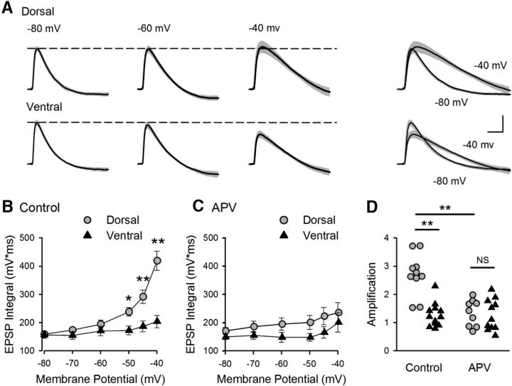
Dorsal-ventral difference in NMDAR-mediated EPSP amplification at SC synapses. A, Average EPSPs recorded from 10 dorsal and 12 ventral cells at the indicated membrane potentials. Shading represents SEM. Right, Traces represent superimposed EPSPs recorded at −80 and −40 mV. Calibration: 2 mV, 20 ms. B, Increase in EPSP integrals with depolarization (EPSP amplification) is significantly smaller in ventral pyramidal cells. *p < 0.05 (two-way ANOVA with Student-Newman-Keuls post hoc multiple-comparisons test). **p < 0.001 (two-way ANOVA with Student-Newman-Keuls post hoc multiple-comparisons test). p = 6.5 × 10⁻⁸, F(1,120) = 33.23. C, EPSP amplification in the presence of the NMDAR blocker d-APV (50 μm) (n = 9 dorsal and n = 10 ventral cells). D, Amplification determined from the ratio of EPSP integrals at −40 and −80 mV for all cells for results shown in B, C. In control recordings, the amplification in dorsal cells (2.7 ± 0.23) was significantly greater than that seen in ventral pyramidal cells (1.3 ± 0.12). Blocking NMDARs significantly reduced amplification in dorsal cells (1.3 ± 0.16) but had no effect on amplification in ventral pyramidal cells (1.3 ± 0.17). **p < 0.001 (two-way ANOVA with Student-Newman-Keuls post hoc multiple-comparisons test). p = 4.9 × 10⁻⁷, F(3,38) = 16.544.

