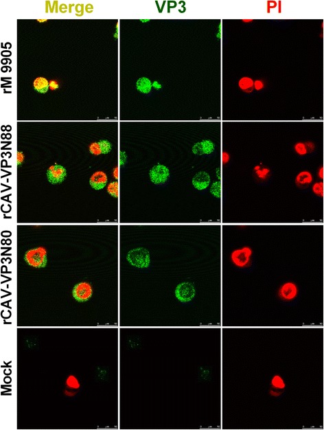
Fig. 5.

Localization of rescued viruses rCAV-VP3N88 and rCAV-VP3N80 compared with parental strain rM9905 in MDCC-MSB1 cells. MDCC-MSB1 cells were infected with rescued virus rCAV-VP3N88 or rCAV-VP3N80 or parental virus rM9905. Mock-infected cells were used as the negative control. Viruses were stained with FITC-conjugated antibody (green) and nuclei with propidium iodide (PI; red). The distribution of apoptin was observed with confocal microscopy. Scale bar is shown at the bottom right (10 μm)
