
Fig. 1.
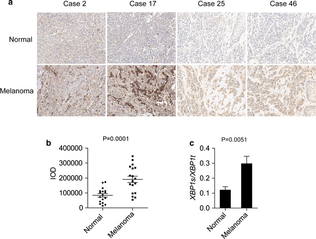
Expression of XBP1s in human melanoma samples. a Representative images and b integrated optical density (IOD) analysis of the immunohistochemical images of melanoma tissues and paired normal skin tissues from 61 melanoma patients. c XBP1 splicing levels were analyzed by real-time PCR in both melanoma tissues and normal skin tissues. The data are presented as the mean ± s.e.m., and P value was analyzed by Student’s t test and labeled