Fig. 1.

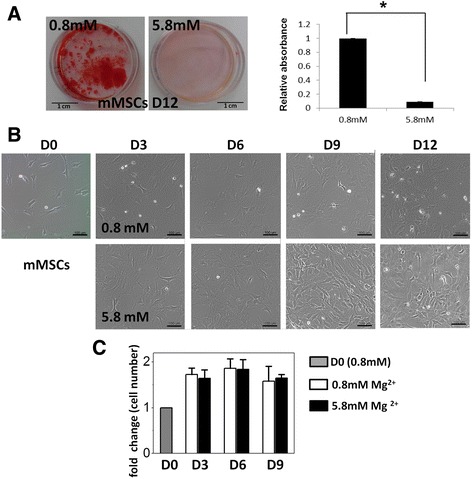
High concentration of extracellular magnesium inhibited mineralization of mouse MSCs during osteogenesis. a Alizarin Red S staining images (left) and quantification (right) of mouse MSCs (mMSCs) 12 days after osteogenic induction with normal (0.8 mM) and high (5.8 mM) extracellular magnesium concentration. Biological replicate N = 3 and technical replicate n = 3 for every biological replicate. b Images of mMSCs during osteogenic differentiation with normal and high extracellular magnesium concentration. Scale bar: 100 μm. c Cell number of differentiating mMSCs under osteogenic induction medium containing 0.8 and 5.8 mM magnesium for 0, 3, 6, and 9 days. Cell number was determined by counting the DAPI-positive cells (N = 3, n = 7) and normalized by cell number for day 0. Data presented as mean ± SEM (* p < 0.05). D: day
