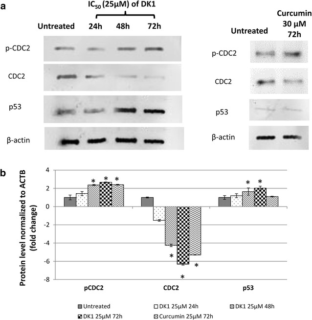
Fig. 5.

Differential protein expression of. a Western blot analysis of CDC2, p-CDC2 and p53 in MCF-7 treated with DK1 (25 μM) for 24, 48 and 72 h. b Differential protein level of control and DK1 (25 μM) treated MCF-7 cells normalised to β-actin. The experiment was done in triplicate and the data are expressed as mean ± SE with *p < 0.05
