
Figure 1.
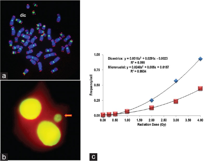
Cytogenetic damage in human lymphocytes following exposure to γ-rays analyzed by chromosome and micronuclei analyses. (a) Peptide nucleic acid-fluorescence in situ hybridization was used to detect dicentric chromosomes. Cy3-telomere (red) and FITC-centromere (green) peptide nucleic acid probes were used along with counterstain DAPI (blue). Metaphase spread shows a dicentric chromosome. (b) Acridine orange stained cytokinesis-blocked binucleated human lymphocytes. Arrow points to a micronucleus present in this cell. (c) Frequencies of dicentrics (blue rectangles) and micronuclei (red rectangles) following gamma irradiation with different doses (0, 0.25, 0.5, 1.0, 2.0, 3.0, and 4 Gy). The data were fitted with polynomial function, and the following equations were obtained: Dicentrics: y = 0.0516x2 + 0.0291x − 0.0023 (R2 =0.999); Micronuclei: y = 0.0249x2 + 0.005x + 0.0157 (R2 = 0.9934)