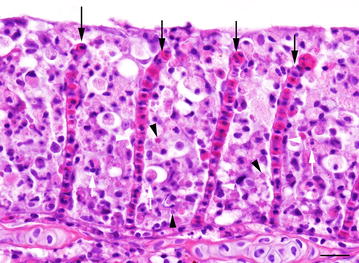
Figure 2.

Gills of KSD affected common carp (donor fish). Clubbing and fusion of secondary gill lamellae with complete occlusion of the interlamellar spaces due to accumulation of cellular debris (white arrows), and hypertrophy of epithelial cells (white arrowheads); Note cytoplasmatic eosinophilic inclusions (black arrowheads); black arrows = lamellar capillaries; HE, bar = 40 µm.
