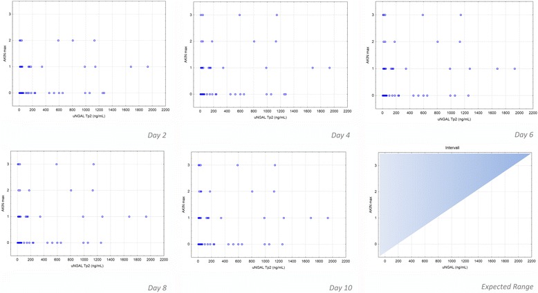
Fig. 3.

Correlation of uNGAL concentrations to corresponding AKIN levels on postoperative days 2, 4, 6, 8, and 10. The scatter-plots show uNGAL values at Tp1 and Tp2. A good prognostic valence is given if the values were mostly in the left upper half of the diagram (shadow area of index chart “expected range”)
