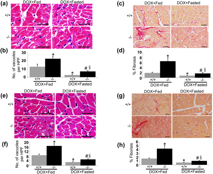
Figure 7. Intermittent fasting ameliorates pathology of DOX-induced cardiotoxicity.
(a) Representative H&E images of degenerative vacuoles in LVs on heart sections from fed or fasted WT and UVRAG-deficient mice 5 days after acute DOX or vehicle treatment. Scale bar: 40 μm. (b) Quantification of degenerative vacuoles in LVs in the experiments as illustrated in (a). n = 3 mice for each group. (c) Representative images of fibrosis stained with picrosirius red in LVs on heart sections from fed or fasted WT and UVRAG-deficient mice 5 days after acute DOX or vehicle treatment. Scale bar: 40 μm. (d) Quantification of fibrosis in LVs in the experiments as illustrated in (c). n = 3 mice for each group. (e) Representative H&E images of LVs on heart sections from fed or fasted WT and UVRAG-deficient mice at 4 weeks of DOX treatment in chronic cardiotoxicity. Scale bar: 40 μm. (f) Quantification of degenerative vacuoles in LVs in the experiments as illustrated in (e). n = 3 mice for each group. (g) Representative images of fibrosis stained with picrosirius red in LVs on heart sections from fed or fasted WT and UVRAG-deficient mice at 4 weeks of DOX treatment in chronic cardiotoxicity. (h) Quantification of fibrosis in LVs in the experiments as illustrated in (g). n = 3 mice for each group. Scale bar: 40 μm. *P < 0.05 vs. WT + DOX + Fed, #P < 0.05 vs. UVRAG−/− + DOX + Fed, §P < 0.05 vs. WT + DOX + Fasted.

