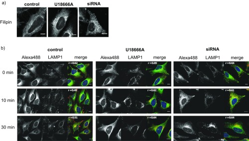
Fig. S7.
(A) Filipin staining of HeLa cell models of NPC. Confocal images of HeLa cells treated with U18666A (2 μg/mL for 24 h) or siRNA against NPC1. Cells were fixed with PFA and stained using Filipin complex (from Staphylococcus aureus, 50 μg/mL). (B) TFS colocalization with endosomes/lysosomes in NPC cell models. Confocal images of HeLa cells treated with U18666A (2 μg/mL for 24 h) or siRNA against NPC1; 6 μM TFS was added for 5 min, and the cells were washed and subsequently uncaged and cross-linked. The time between uncaging and cross-linking was varied from 0 min to 30 min. Cells were fixed and washed, and the cross-linked lipids were functionalized with Alexa488. Late endosomes/lysosomes are visualized via LAMP1 antibody in gray (middle image) and red (merged image), and Pearson’s correlation coefficient is shown in the top left corner of the merged image. (Scale bars, 20 μm.)

