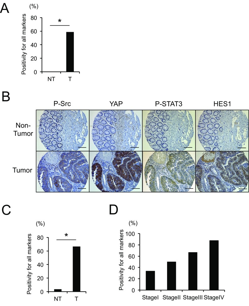
Fig. S1.
(A) Human nontumor (NT) and tumor (T) colon tissue specimens were stained with P-Src, YAP, P-STAT3, or HES1 antibodies. The percentage of tumors positive for all four markers is shown. *P < 0.05. (B and C) TMA of human CRC (n = 27) and matched normal colon sections (n = 27) were stained with P-Src, YAP, P-STAT3, or HES1 antibodies. Representative examples of one nontumor and one tumor sample are shown (B). (Scale bars, 100 μm.) The percentage of tumors positive for all four markers was determined (C). *P < 0.05. (D) The percentage of tumors positive for all four markers in each tumor-node-metastasis (TNM) stage is shown.

