Fig. 1.
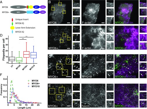
Expression of MYO6+ leads to formation of elongated filopodia. (A) Constructs used in this study: MD, motor domain; MT, medial tail; CBD, cargo binding domain. (B) TIRF images of HeLa cells expressing either GFP-MYO6 (Top) or GFP-MYO6+ (Bottom) showing formation of filopodia, which extend across the coverslip surface. Insets shown are 8× magnification. (C) TIRF image showing colocalization of MYO6+ and MYO10 at the tips of filopodia. Insets shown are 4× magnification. (D) Box-and-whisker plot of the number of filopodia in cells either untransfected or expressing the indicated GFP-tagged constructs (more than 60 cells per construct over three independent experiments). ***P < 0.001. (E) TIRF image showing MYO6+ at the tips of fascin bundles. Insets shown are 4.5× magnification. (F) Normalized distribution of filopodia length in cells expressing GFP-MYO6 (black circles, 149 cells, three experiments), GFP-MYO6+ (blue circles, 233 cells, three experiments), or GFP-MYO10 (red circles, 180 cells, three experiments). Errors shown are SEM. (Scale bars, 10 µm.)

