Fig. 3.
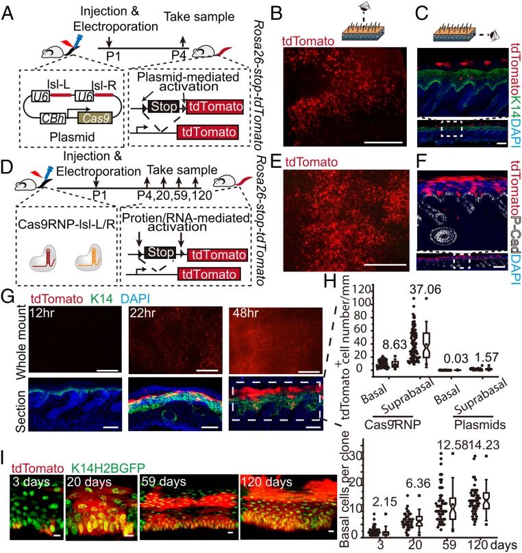
Efficient in vivo deliveries of Cas9/sgRNA ribonucleoproteins into skin stem cells of postnatal mice using electroporation. (A) Schematic diagram of electroporation with pSpCas9-lsl-L/R plasmid at the tail of Rosa26-stop-tdTomato reporter newborn mice. Representative whole mount (B) and section (C) immunofluorescence staining of Rosa26-stop-tdTomato mouse tail skin after pSpCas9-lsl-L/R plasmid electroporation. (Scale bar, 1 mm for whole mount images, 100 μm for section staining images.) (D) Schematic diagram of electroporation with Cas9/sgRNA ribonucleoproteins at the tails of Rosa26-stop-tdTomato reporter newborn mice. Representative whole mount (E) and section (F) immunofluorescence staining of Rosa26-stop-tdTomato reporter tail skin at 3 d after treatment. (Scale bar, 1 mm for whole mount images, 500 μm for section staining images.) (G) Representative whole mount and section immunofluorescence stainings of Rosa26-stop-tdTomato reporter tail skin at 12 h, 22 h, and 48 h post-Cas9/sgRNA ribonucleoprotein electroporation. (Scale bar, 500 μm for whole mount images, 70 μm for section images.) (H) Quantifications of the number of tdTomato+ cells in the basal and suprabasal layers of epidermis at 3 d postelectroporation treatments with either ribonucleoproteins or plasmid DNA. (I) Expansion of basal cell per colony after one electroporation treatment with Cas9/sgRNA ribonucleoproteins. Representative images at 3, 20, 59, and 120 d posttreatment are shown at Left; quantifications are shown at Right. (Scale bar, 10 μm.)

