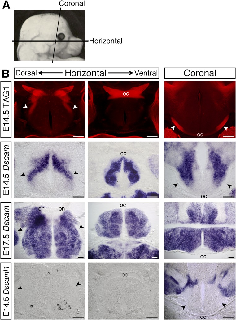
Fig. S1.
Expression of Dscam along the developing optic pathway. (A) Schematic illustration of the plains of sections used. (B) Serial horizontal sections through the ventral diencephalon and coronal sections at the level of the optic tracts of E14.5 and E17.5 C57BL/6J embryos stained with antibodies against TAG-1 to label the RGC axons (Top), or by in situ hybridization with probes specific for Dscam or Dscaml1. Horizontal sections, anterior up; coronal sections, dorsal up. oc, optic chiasm; on, optic nerve; arrowheads, optic tracts. (Scale bars: 200 µm.)

