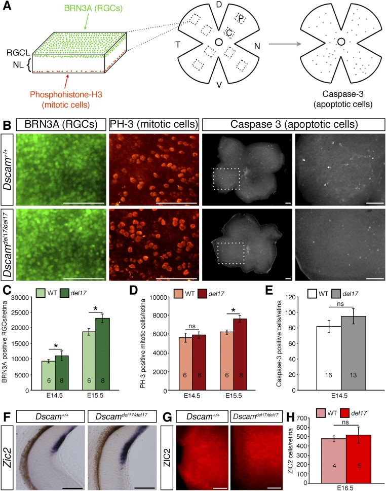
Fig. S4.
RGC genesis is increased in Dscamdel17 mutants, but specification of ipsilaterally projecting RGCs occurs normally. (A) Schematic illustration of the method used to quantify RGC, mitotic, and apoptotic cell numbers. Retinas were stained by double immunofluorescence with antibodies against BRN3A to label RGCs, with phosphohistone H to label mitotic cells, or with cleaved caspase-3 to label apoptotic cells. Confocal images were captured through the RGC, and neuroblastic layers from four peripheral (p) and four central (c) regions in each retina of BRN3A- and phosphohistone H3-positive cells, and the number of labeled cells in each image was quantified and used to calculate the total number of labeled cells in each retina. Caspase-3–positive cells were quantified by counting all labeled cells. D, dorsal; N, nasal; NL, neuroblastic layer; RGCL, RGC layer; T, temporal; V, ventral. (B) Confocal images of BRN3A-positive RGCs and phosphohistone H3-positive mitotic cells in the RGC layer and neuroblastic layer respectively, and caspase-3–positive cells in E14.5 Dscamdel17 WT and mutant retinas. (C–E) Mean ± SEM number of BRN3A-positive RGCs (C), phosphohistone-H3–positive mitotic cells (D), and caspase-3–positive apoptotic cells (E) in retinas from E14.5 and E15.5 Dscamdel17 WT and mutant (del17) retinas. (F) Sections of E15.5 Dscamdel17 WT and mutant retinas stained by in situ hybridization with a probe specific for Zic2, a marker of ipsilaterally specified RGCs. (G) Peripheral ventrotemporal retina from E16.5 Dscamdel17 WT and mutant littermates stained with an antibody against ZIC-2. (H) Mean ± SEM number of ZIC2-positive cells in retinas from E16.5 Dscamdel17 mutant (del17) and WT littermates. *P < 0.05; ns, not significant. The numbers analyzed at each age and genotype are indicated on the bars. (Scale bars: 100 µm.)

