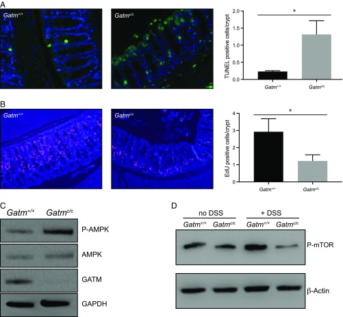
Fig. 5.
Colonic epithelium of Gatmc/c mice exhibit reduced proliferation and increased cell death during DSS treatment. (A) Images of TUNEL staining (green) of colon sections from Gatm+/+ and Gatmc/c mice on day 4 of 1.5% DSS treatment (20× magnification); images are representative of three independent experiments. Quantification was performed on a minimum of 50 crypts for Gatm+/+ (n = 3) and Gatmc/c (n = 3) colons. *P < 0.05. (B) Metabolic labeling of DNA in Gatm+/+ and Gatmc/c colons on day 4 of 1.5% DSS treatment. EdU-labeled DNA was stained with Click-iT EdU labeling Alexa Fluor 647 (20× magnification); images are representative of at least three independent experiments. Quantification was performed on a minimum of 50 crypts for Gatm+/+ (n = 5) and Gatmc/c (n = 5) colons. *P < 0.05. (C and D) Colonic epithelium extracts were assessed by Western blot on day 4 of 1.5% DSS treatment for phosphorylated AMPK (C) and phosphorylated mTOR (D). Results are representative of more than three independent experiments with at least three mice per group. (A and B) P values were calculated by Student’s t test; bars indicate mean ± SD.

