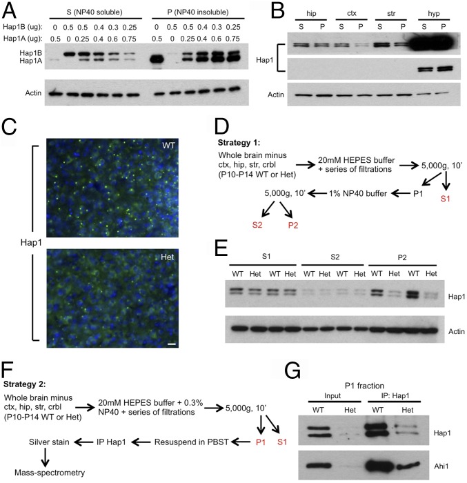
Fig. 1.
A strategy to isolate the SB-enriched fraction from mouse brain. (A) Different ratios of Hap1A to Hap1B were transfected into HEK293 cells, and the lysate was separated into Nonidet P-40–soluble (S) and Nonidet P-40–insoluble (P) fractions. Western blotting shows that, as the ratio of Hap1A to Hap1B increases, more Hap1 is found in the P fraction. (B) Western blotting of Hap1 on S and P fractions from the hippocampal (hip), cortical (ctx), striatal (str), and hypothalamic (hyp) tissues isolated from postnatal day 18 WT mouse brain. Both longer (Upper) and shorter (Middle) exposures for Hap1 are shown. (C) Immunostaining of Hap1 in postnatal day 14 WT and Hap1-KO (Het) mouse hypothalamus demonstrates markedly fewer Hap1 puncta in Het hypothalamus. (Scale bar: 20 µm.) (D) A strategy to isolate the SB-enriched fraction. (E) Western blotting of Hap1 on WT and Het mouse brain fractions obtained using strategy 1. (F) A strategy to use the SB-enriched fraction for identifying novel proteins associated with Hap1-contianing SB. (G) IP of Hap1 in P1 fractions from postnatal day 11 WT and Het mouse brain derived with strategy 2.

