Figure 7.

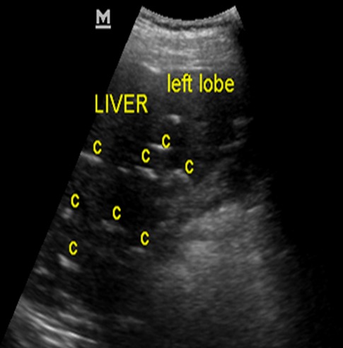
Abdominal ultrasonogram of case 2 at the level of the liver showing multiple brightly echogenic lesions (C) casting posterior acoustic shadows in the liver parenchyma, distorting its architecture

Abdominal ultrasonogram of case 2 at the level of the liver showing multiple brightly echogenic lesions (C) casting posterior acoustic shadows in the liver parenchyma, distorting its architecture