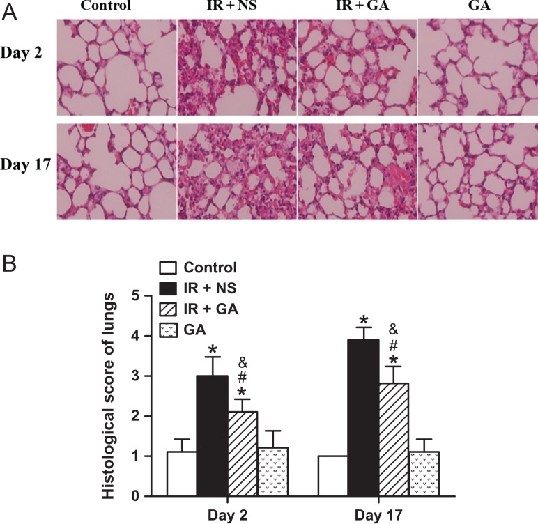
Fig. 1.
Histologic detection and semi-quantification of radiation-induced lung injury in mice. Mice of the IR + NS group and IR + GA group were exposed to a single fraction of X-ray irradiation (12 Gy) to the thorax and administered NS or GA. The GA group was not exposed to irradiation. Panel A: Typical hematoxylin and eosin (H&E)-stained sections of mouse lungs from each group 2 days and 17 days after irradiation (magnification × 400). Panel B: Histologic scores of lung injury from H&E-stained sections. Data are the mean ± SD. *P < 0.05, vs control group; #P < 0.05, vs IR + NS group; &P < 0.05, vs GA group, at identical time-points, by one-way ANOVA (followed by Bonferroni's multiple comparison test). IR = irradiation, NS = normal saline, GA = glycyrrhetinic acid.

