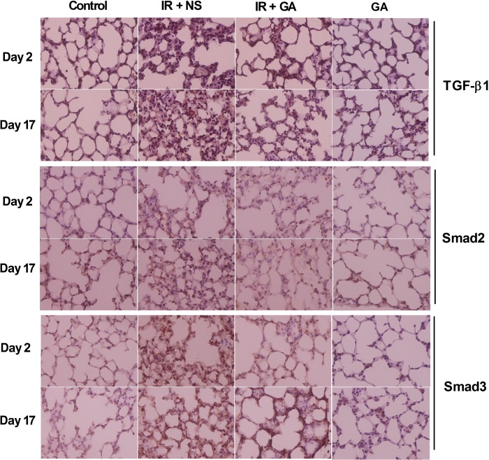
Fig. 4.
Immunohistochemical staining of mouse lung tissue after thoracic irradiation. Mice in group IR + NS and IR + GA were exposed to a single fraction of 12 Gy and administered NS or GA. Mice in group GA were not irradiated. Typical TGF-β1-, Smad2- or Smad3-stained sections of mouse lungs from each group 2 days and 17 days after irradiation (×400 magnification). IR = irradiation, NS = normal saline, GA = glycyrrhetinic acid.

