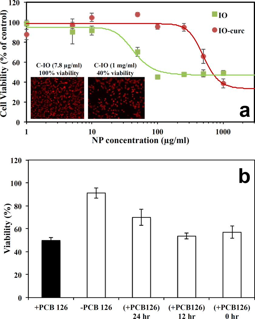
Fig. 4.
Shows the cell viability assay analysis using C-IO MNPs. Part a) Dose dependent exposure of uncoated iron oxide and curcumin-iron oxide nanoparticles towards HUVECs for 24 hours followed by viability analysis using Calcein AM red-orange live cell tracer. Inset: Fluorescent images of the HUVECs after 24-hour exposure followed by calcein AM red-orange dying. Part b) Protection against PCB 126 induced inflammation. HUVECs preincubated with 10 µg/mL curcumin iron oxide nanoparticles for 0, 12 and 24 hours followed by 24-hour exposure to 50 µM PCB126.

