Fig. 5.
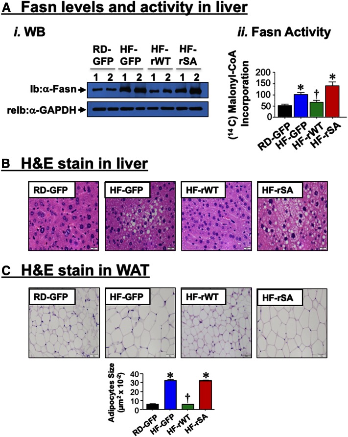
Effect of adenoviral-mediated redelivery of CEACAM1 on diet-induced lipid alterations. A-i: Western analysis of Fasn levels in the liver lysates from mice fed with RD or HF for 41 days and injected with adenoviral particles in the last 21 days of feeding. ii: Fasn activity was assayed in liver lysates of RD-GFP (black), HF-GFP (blue), HF-rWT (green), or HF-rSA (red) (n = 6–8/per group). Values are expressed as mean ± SEM. * P < 0.05 versus RD-GFP; † P < 0.05 versus. HF-GFP. B: Liver histology was assessed by H and E stained sections (n = 5/each group). Whereas HF-GFP and HF-rSA exhibited microvesicular lipid infiltration alternating with normal liver parenchyma, RD-GFP and HF-rWT exhibited normal histology. Representative images from three sections/mouse are shown (40× magnification). C: H and E stain on WAT sections (n = 5/each group). HF diet caused enlarged adipocytes relative to RD-fed mice (HF-GFP vs. RD-GFP), as shown by the adipocyte size in the accompanying bar graph. This was reversed by Ad-rWT, but not Ad-rSA, injection. Representative images from three sections/mouse are shown (20× magnification), and values of adipocyte size are expressed as mean ± SEM. * P < 0.05 versus RD-GFP; † P < 0.05 versus HF-GFP.

