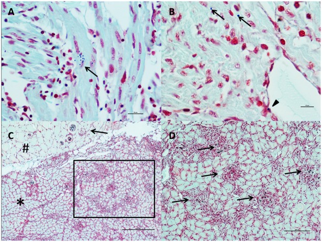
Fig 6. P. theridion and K. thyrsites detection in the tissues through Twort’s Gram staining.
A) Cluster of P. theridion (small, dark blue organisms) inside myocardial cells in the spongy layer (arrow). Bar scale: 20μm. B) P. theridion in myocardial cells (arrows) and in an endocardial cell (arrowhead). Scale bar: 10μm. C) K. thyrsites inducing nodular granulomatous myositis in red muscle. The parasites are located in both red (star) and white (pound) portion of the skeletal muscle. One plasmodium inside a white muscle cell is also inducing a nodular granulomatous inflammatory reaction (arrow). Scale bar: 250μm. D) Inset of Fig C. Dark blue parasites are visible inside the nodular inflammation sites: 100μm.

