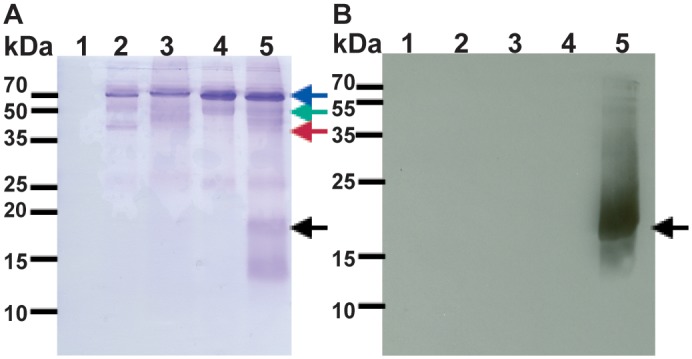
Fig 11. Co-immunoprecipitation of PelB-Vpu with CD4.

Purified CD4 TMD + cytoplasmic domain fused to the amino end of MBP (CD4-MBP) was incubated with PelB-Vpu in βDDM micelles. The complex was captured by anti- MBP antibody-conjugated beads and pelleted by centrifugation and analyzed by SDS-PAGE followed by Coomassie staining (A) and immunoblot analysis with Vpu antibodies (B). Panel A depicts all proteins pulled down by only agarose beads (lane 1) and agarose beads conjugated with anti-MBP (lanes 2–5). Anti-MBP, CD4-MBP, MBP and Vpu are denoted by blue, green, red and black arrows respectively. Beads were incubated with purified proteins: CD4-MBP (lane 1), MBP and Vpu (lane 2), CD4-MBP (lane 3), Vpu lane 4), CD4-MBP and Vpu (lane 5).
