Figure 1.
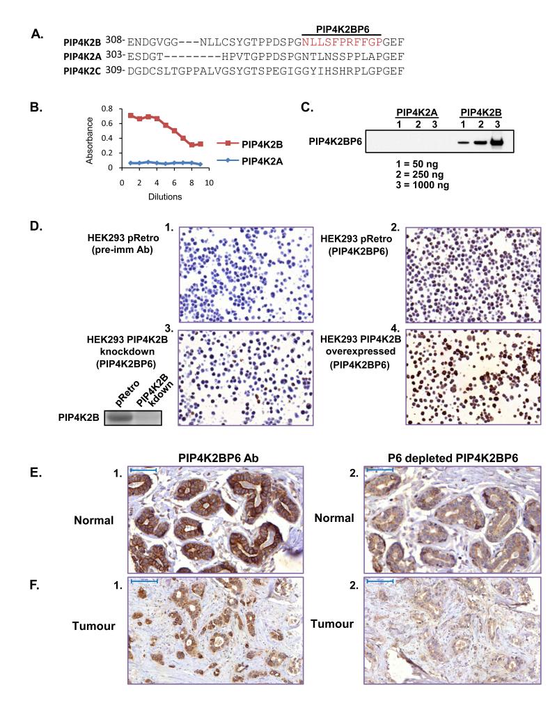
Characterisation of the PIP4K2BP6 antibody. A. Sequence alignment of the region surrounding the epitope (in red) for PIP4K2BP6 in PIP4K2A, PIP4K2B and PIP4K2C. B. ELISA of serial dilutions of PIP4K2BP6 against recombinant PIP4K2B and PIP4K2A protein. C. Western blot of recombinant PIP4K2B and PIP4K2A using PIP4K2BP6. D. HEK293 cells were transduced with a control vector (pRetro panels 1 and 2), or a shRNA construct targeting PIP4K2B (panel 3). The shRNA vector targeting PIP4K2B decreased PIP4K2B protein levels compared to control vector (inset next to panel 3). Cells were also transduced with a construct to overexpress PIP4K2B (panel 4). Cells were fixed and embedded in paraffin and stained with a control pre-immune antibody (panel 1) or the PIP4K2BP6 antibody (panels 2, 3 and 4). The images show that PIP4K2BP6 specifically recognises PIP4K2B. E, F. Normal breast tissue (E) or tumour tissue (F) was stained with PIP4K2BP6 before (panel 1) or after (panel 2) specific depletion of PIP4K2BP6 with the P6 peptide. Scale bars represent 50μm (E) or 100μm (F)

