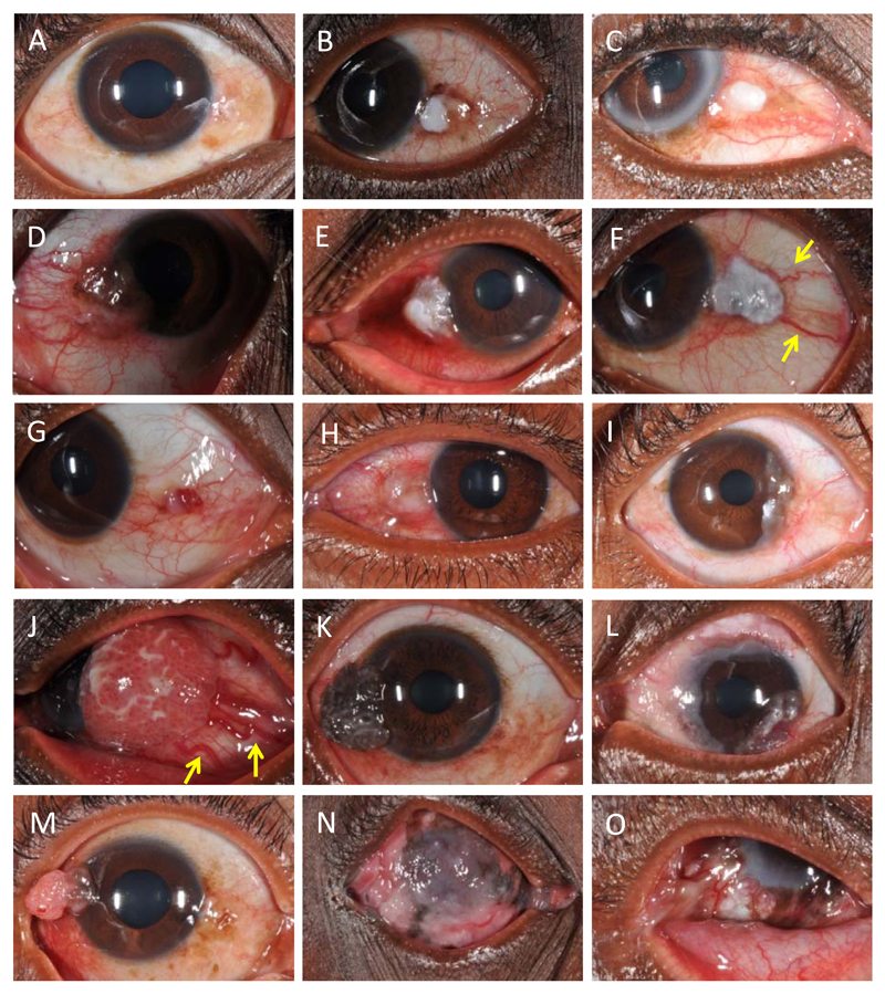
Figure 1. Five grades of inflammation associated with OSSN are shown in A-E. Various clinical features seen in moderately differentiated squamous cell carcinoma are shown from F to O. F-K shows different tumour surface appearances and various growth patterns are seen in L-O.
(A) No inflammation; (B) Minimal inflammation with leukoplakia and brown pigmentation;n (C) Mild inflammation with leukoplakia; (D) Moderate inflammation with leukoplakia; (E) Severe inflammation with leukoplakia; (F) Leukoplakia – patches of keratosis visible as white adherent plaques. Feeder vessels (distinctly dilated blood vessels larger than the rest of conjunctival vessels) are also shown by yellow arrows; (G) Erythoplakia – a red subconjunctival popular haemorrhage-like appearance; (H) Gelatinous appearance; (I) Fibrovascular appearance; (J) Papilliform appearance with markedly large feeder vessels (yellow arrows); (K) Brown pigmentation; (L) circumlimbal lesion; (M) pedunculated lesion;(N) Extensive corneal involvement with orbital disease; (O) Symblepharon.

