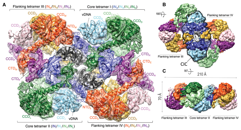
Fig. 1. Cryo-EM reconstruction of the MVV intasome.
A. Fitted intasome model color-coded to highlight IN subunits including 12 NTDs, 16 CCDs, and 14 CTDs. Molecules of vDNA in dark grey are surrounded by core tetramers I and II (colored in green, light green, sky blue, and blue), and flanking tetramers III and IV (red, yellow, pink, and purple). B-C. Views of the map in two alternative orientations. The CIC structure is highlighted with a black outline in panel B.

