
Figure 1.
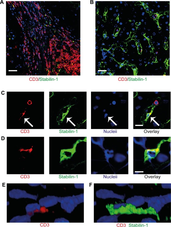
Lymphocytes migrate into sinusoidal endothelial cells in human chronic liver disease. (A) Immunofluorescent staining of liver sections from a patient with primary biliary cirrhosis demonstrating the presence of lymphocytes in portal regions within fibrous septum. (B) Immunofluorescent staining of the same liver demonstrating lymphocytes within the sinusoidal channels. CD3+ lymphocytes appear in red and stabilin‐1–positive hepatic sinusoidal endothelium appears in green. (C) Lymphocytes (red) within the sinusoidal channels with one lymphocyte within the sinusoidal lumen and another within the endothelial cell (green). Arrows indicate the intraendothelial lymphocyte. (D) Two‐dimensional image of orthogonal (XZ) projection of a CD3+ lymphocyte (red) colocalizing with a stabilin‐1–positive (green) endothelial cell. (E,F) Three‐dimensional reconstruction of the orthogonal (XZ) projection in panel D with CD3+ red signal only (E) and overlay of stabilin‐1–positive green signal (F). Scale bars = 20 μm (A,B), 10 μm (C), and 5 μm (D).