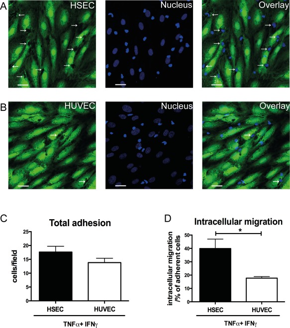
Figure 4.

IFNγ promotes intracellular migration of lymphocytes into HSECs but not HUVECs. (A,B) Representative confocal images of lymphocytes adherent to TNFα‐ and IFNγ‐treated HSEC and HUVEC monolayers in a flow adhesion assay. Endothelial cells were stained with CellTracker CFMDA (green) and nuclei were stained with DAPI (blue). Arrows indicate intracellular lymphocytes. (C) Quantification of adhesion and (D) intracellular migration of lymphocytes into HSEC and HUVEC monolayers in a flow adhesion assay. Quantitative data are the mean ± SEM of five independent experiments. Statistical significance was determined using a two‐tailed t test. *P < 0.05. Scale bars = 25 μm (A,B).
