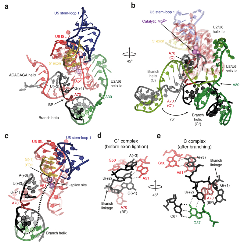
Figure 2. Architecture of the RNA catalytic core in C* complex.
a, Key RNA structures at the active site. The branch helix has undocked from the catalytic Mg2+ site. BP, branch point; ISL, internal stem-loop; M1 and M2, catalytic metal ions. b, Rotated view showing superposition of the RNA catalytic core for the C (PDB, 5LJ5; ref. 4) and C* spliceosomes. C elements are coloured in light shades. Note substantial rotation of the branch helix between the two complexes. c, The BP and 5’-splice site nucleotides align in a path to the catalytic Mg2+ site. A possible intron path guiding the 3’-splice site to the Mg2+ site is shown as a dashed line. d, Watson-Crick-Hoogsteen interaction between U(+2) and A51 of U6 snRNA. e, Different interactions of U(+2) and A(+3) observed in C complex.

