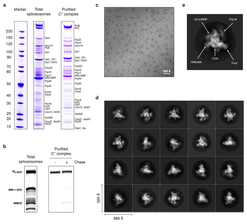
Extended Data Figure 1. Purification and Cryo-EM imaging of the C* spliceosome.
a, Protein composition of the purified C* complex. Note that Prp16 is significantly de-enriched compared to Prp22, consistent with the majority of the purified complexes being in a post-Prp16 conformation, as Prp16 dissociates upon ATP hydrolysis8. b, The purified C* complex contains mostly lariat-intermediate and catalyses exon ligation with low efficiency when incubated in the presence of Mg2+. The identity of the major species, inferred by size and migration pattern, is indicated by cartoon on the left. c, Representative electron micrograph of the C* complex sample collected at 3 μm defocus. d, Representative 2D class averages of the C* complex obtained in RELION39. e, Image of a highly abundant C* complex 2D class average illustrating the major domains of the complex.

