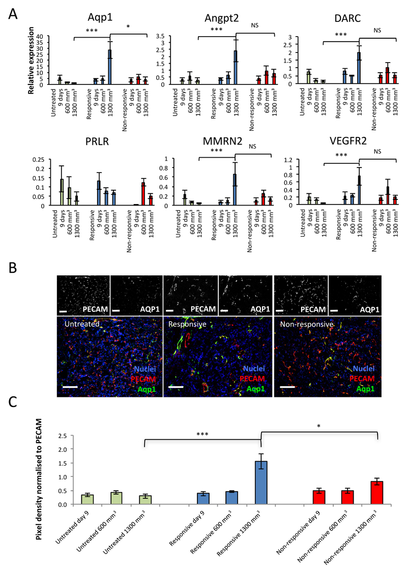
Figure 4. Aquaporin is significantly enriched in the vessels of responsive tumours over those of untreated and non-responsive tumours.
A, RTqPCR for the relative expression of the six genes of interest in endothelial isolates from untreated, responsive and non-responsive tumours harvested at 9 days, 600 mm3 and 1300 mm3 (mean expression ±SEM, *** p<0.001, * p<0.05, NS – Not Significant, Mann-Whitney). B, representative images of AQP1 staining in untreated, responsive and non-responsive tumours by immunofluorescence (IF). Black and white split channel and colour merged channel images of tumours triple stained by IF for DAPI (nuclei, blue), PECAM-1 (vessels, red) and AQP1 (green). C, quantitation of pixel density of staining by IF for AQP1 standardised to PECAM-1 staining (mean ± SEM, *** p<0.001, * p<0.05, Mann-Whitney, n-numbers: Day 9, responsive (R)=5, non-responsive (NR)=5, untreated (UT)=10; 600mm3, R=4, NR=6, UT=10; 1300mm3, R=12, NR=7, UT=17, 10 fields of view each).

