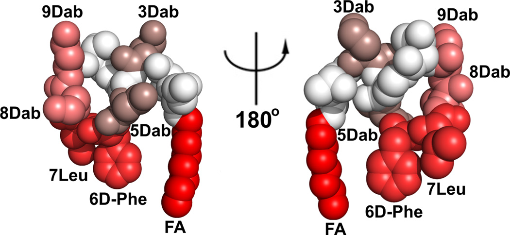
Figure 4.
Monoclonal antibiody clone 45 epitope ‘HOTSPOTS’ on the polymyxin B structure. The SPR KD values were mapped onto the NMR solution structure of polymyxin B1 (higher affinity binding is represented by a brighter red coloring). The polymyxin B1 structure is shown in CPK representation and the two views are rotated by 180° about the y-axis.

