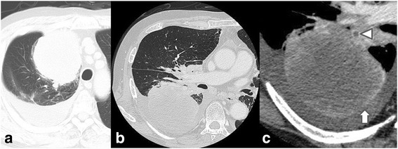
Fig. 2.

a Plain CT at first visit showing a large tumor mainly located at right S1-3, widely in contact with the precordial mediastinum and pleura. Massive pleural effusion in the right thoracic cavity is also present. Contrast-enhanced CT image at the onset of torsion, b lung window, c mediastinal window (enlarged image). The tumor in the right upper lung lobe has moved to the posterior thoracic space. There is a thrombus in the upper pulmonary vein (triangle) and non-contrast regions in the peripheral tumor (upward arrow), leading to suspicion of blood flow impediment
