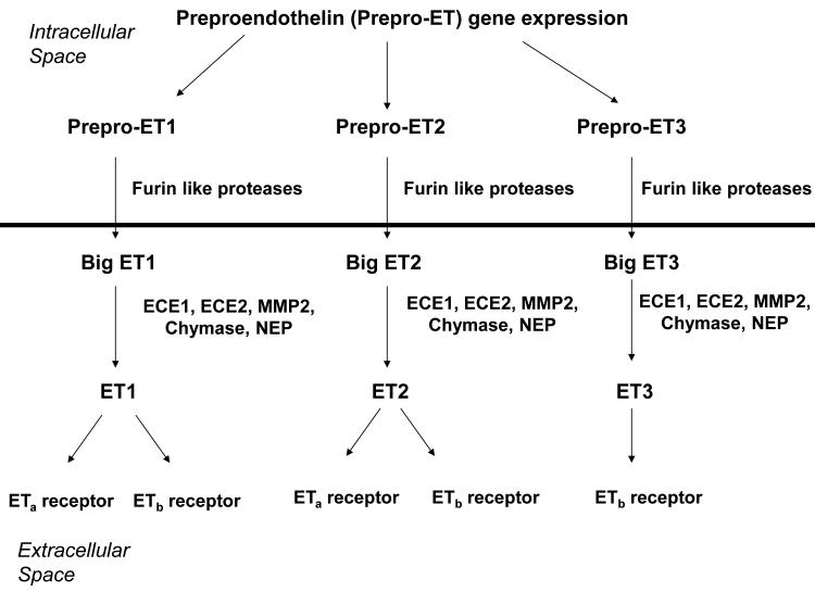
Figure 1. Schematic representation of endothelin synthesis, secretion, and receptor binding.
The horizontal line represents the cell membrane, with events above the line occurring intracellularly and those below the line occurring extracellularly. Both the ET receptors and the ECEs are membrane bound, but their ligand binding/catalytic sites are extracellular. ETA represents the A type endothelin receptor, encoded by EDNRA in humans and Ednra in mice. ETb represents the B type endothelin receptor, encoded by EDNRB in humans and Ednrb in mice. All 3 endothelins are initially synthesized as pre-propeptides, encoded by EDN1, EDN2, and EDN3 in humans and Edn1, Edn2, and Edn3 in mice. They are processed to the respective big ETs by furin-like proteases prior to secretion. The big ETs are further processed to the mature, biologically active ETs by the ECEs or other extracellular proteases. B type receptors in endothelial cells promote NO synthesis, cell survival, and ET clearance. Both A type and B type receptors promote smooth muscle cell contraction and collagen synthesis by fibroblasts.

