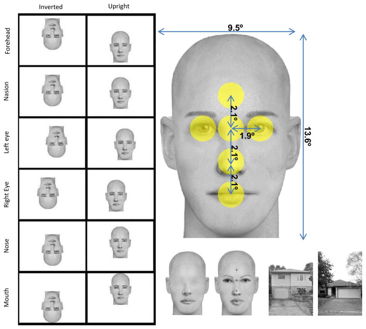
Figure 1.
Left panel: examples of one face presented at each fixation location in the inverted and upright conditions. Participants fixated in the center of the monitor represented here by each white rectangle (images are to scale) and the face was offset in such a way that gaze fixated 6 possible face locations: forehead, nasion, left eye, right eye, tip of the nose, and mouth. Note that eye positions are from a viewer perspective (i.e. left eye is on the left of the picture). This particular positioning of the face to obtain the desired feature fixated resulted in the face situated almost entirely in the upper visual field when fixation was on the mouth in upright faces and almost entirely in the lower visual field when fixation was on the forehead, while the opposite pattern was seen for inverted faces. Right panel, up: one face exemplar with picture size and distances between features in visual angles. The yellow circles represent the interest areas of 1.8° centered on each feature that were used to reject eye gaze deviations in each fixation condition. Right panel, bottom: houses, eyeless and intact face examples, with fixation crosses overlaid on the intact face to indicate where the fixation would occur (note that fixation crosses were never presented overlaid on faces in the actual experiment).

