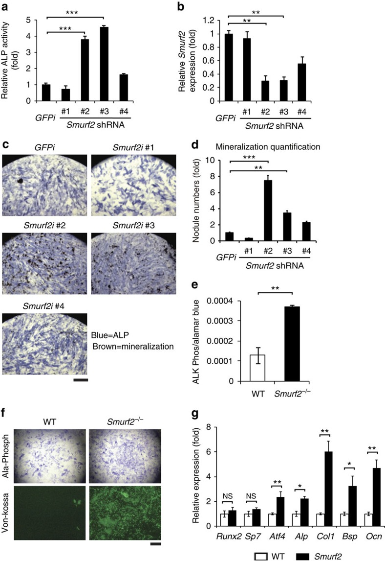
Figure 1. Osteoblast differentiation increased in Smurf2 knockdown and knockout cells.
(a,b) Analysis of ALP and Smurf2 expression in human mesenchymal stem cells (hMSCs) infected with lentivirus-expressing GFP control or Smurf2 shRNAs. (c) Von Kossa staining representative images for combined ALP (blue) and Von Kossa (brown) staining of hMSCs infected with lentivirus-expressing control or Smurf2 shRNAs cultured in osteoblast differentiation medium for 21 days. Scale bar, 0.1 cm. (d) Bone nodule mineralization of hMSCs determined by Von Kossa staining. The bar graph displays the quantification of Von Kossa-positive mineralized nodules. (e,f) Analysis of ALP expression and Von Kossa staining of primary osteoblasts cultured in osteoblast differentiation medium for 7 and 21 days. Scale bar, 0.1 cm. (g) Quantitative reverse transcriptase–PCR analysis of osteoblast genes in primary osteoblasts from neonatal 5-day-old mice cultured in osteoblast differentiation medium for 7 days. Data represent mean±s.d. (n=10 for each genotype, *P<0.05, **P<0.01, ***P<0.001, NS, not significant. Student's t-test).

