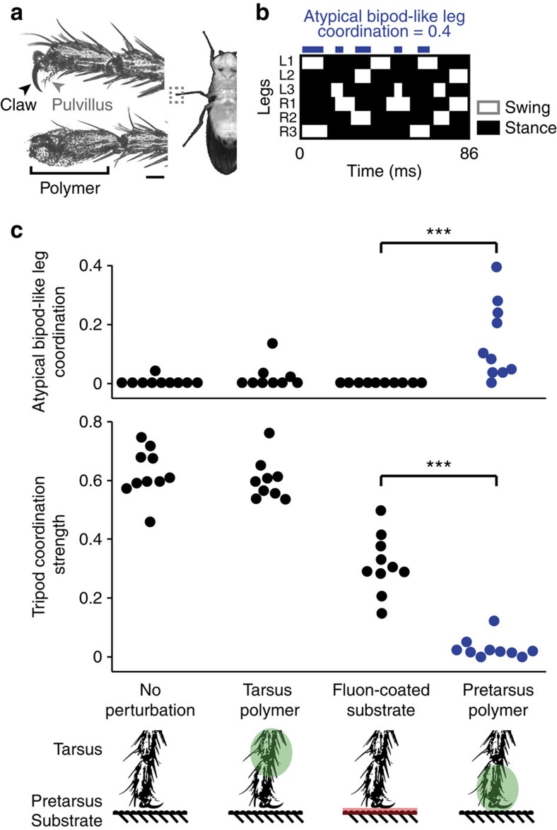
Figure 6. Blocking leg adhesion in D. melanogaster abolishes the tripod gait and uncovers the potential for atypical bipod-like leg coordination.
(a) The pretarsus, the distal-most segment of the D. melanogaster leg (grey dashed box, right), houses a claw (black arrowhead) and pulvillus attachment pad (grey arrowhead), which are used to adhere to surfaces (left, top). We used a ultraviolet-curing polymer to cover pretarsal adhesive structures (left, bottom). Scale bar, 40 μm. (b) Footfall diagram for a fly walking with polymer coating on each pretarsus. Contact with the ground during stance phase (black) and no ground contact during swing phase (white) are indicated for each leg over time. Blue blocks indicate periods of atypical bipod-like leg coordination. This animal exhibits atypical bipod-like leg coordination 40% of the time. (c) Atypical bipod-like leg coordination (top) and TCS (bottom) for unperturbed flies (left), flies with polymer on each tarsus (green, middle-left), flies walking on a Fluon-coated substrate (pink, middle-right) or flies with polymer on each pretarsus (green, right). N=10, 9, 10 and 10 flies, respectively. A triple asterisk (***) indicates that P<0.001 for a Wilcoxon's rank-sum test.

