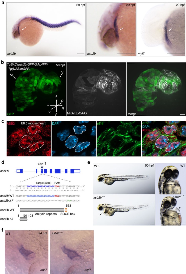
Figure 1. asb2b mutant zebrafish exhibit cardiac defects.
(a) In situ hybridization for asb2b and myl7 expression in 29 hpf embryos. asb2b is expressed in the myl7+ heart tube (arrows) and in the somites. Lateral views, anterior to the left. Scale bars, 200 μm. (b) Three-dimensional images of 50 hpf Tg(asb2b:GFP-GAL4FF);Tg(UAS:mGFP);Tg(myl7:MKATE-CAAX) hearts. Scale bar, 40 μm. (c) E8.5 mouse heart co-stained for ASB2 (red), cardiac troponin I (cTnI, green) and 4,6-diamidino-2-phenylindole (DAPI; blue), showing nuclear and cytoplasmic localization of ASB2; dashed lines outline nuclei. Scale bars, 10 μm. (d) Schematic representation of the asb2b locus. The gRNA target sequence, PAM and premature stop codon are highlighted in blue, red and green, respectively. Deleted nucleotides are indicated by dashes. Predicted structure of WT and mutant (Asb2b Δ7) proteins. The asb2b Δ7 allele (asb2bbns33) is predicted to encode a truncated polypeptide containing two incorrect amino acids (102–103 aa). (e) Bright-field micrographs of 50 hpf WT and asb2b mutant embryos in lateral and ventral views. Black arrows point to pericardial edema. Scale bars, 200 μm (f) in situ hybridization for myl7 expression in 24 hpf WT and asb2b mutant. Scale bar, 100 μm. At, atrium; A, anterior; D, dorsal; L, left; P, posterior; R, right; V, ventricle; V, ventral.

