Figure 2.

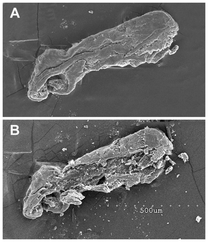
Figure 2 A. A scanning electron micrograph of the specimen from case 1 demonstrating the endolymphatic duct with contained particulate matter. Figure 2 B. The same specimen after the exposed surface layer (presumably the endolymphatic duct wall) was mechanically removed exposing the contents of the canal (particulate matter). Scale bar is shown in panel B 500μm
