
Figure 2. Morphological and Proliferative Differences Between LTS-Scar and Normal Human Laryngotracheal Fibroblasts.
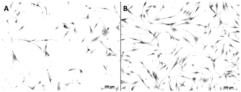
Normal (A) and LTS-scar (B) were imaged at Day 6 at 10× magnification. Scar cells demonstrated an increased cell surface area compared with normal cells (p < 0.05).