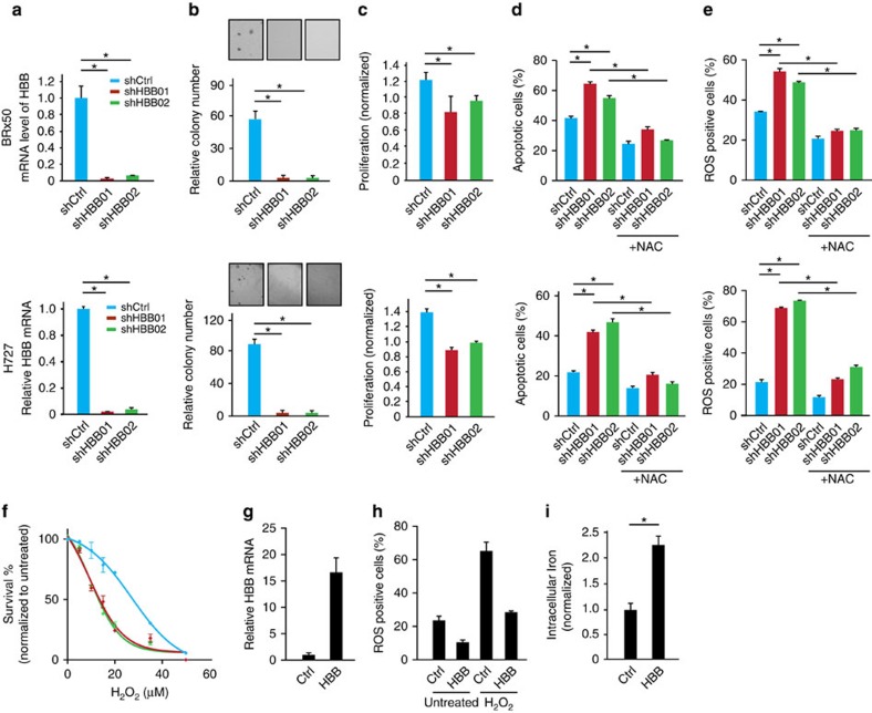
Figure 3. HBB contributes to the survival of tumour cells under ROS stress.
(a–e) top panel: BRx50 cells; bottom panel: H727 cells (a) Bar graph showing effective shRNA-mediated knockdown of HBB. (b) Bar graph showing that HBB depletion impairs colony formation in soft agar (measured at 3 weeks). Representative images are shown. (c) Bar graph showing that depletion of HBB reduces short-term proliferation of BRx50 cells (5 days) and H727 cells (24 h) cultured in suspension. (d,e) Bar graph showing that HBB depletion increases apoptosis and intracellular ROS levels in cells cultured in suspension for 24 h; pre-treatment of cells with the anti-oxidant NAC, rescues both ROS levels and apoptosis. (f) Bar graph showing that HBB-depleted H727 cells exhibit increased sensitivity to H2O2 compared with control cells. (g) Real-time PCR showing the relative HBB mRNA levels in BRx50 cells at baseline (Ctrl) or following stable expression of ectopic HBB. (h) Bar graph showing that ectopic overexpression of HBB in BRx50 cells reduces intracellular ROS under basal conditions and following treatment with hydrogen peroxide. (i) Bar graph showing that overexpression of HBB increases total iron within BRx50 cells compared with control cells. All data (a–i) are represented as mean±s.d. n=3; *denotes a statistical significance at P<0.05 (t-Test).

