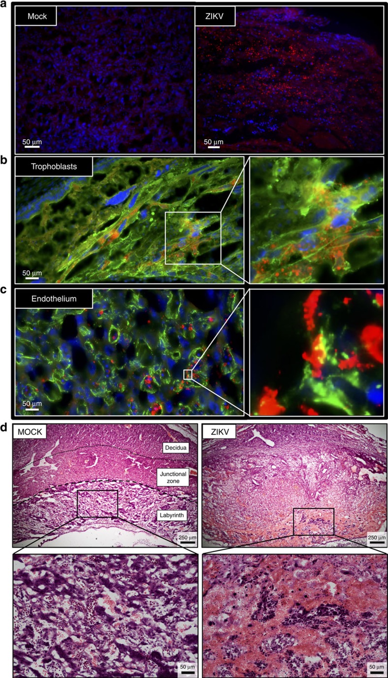
Figure 4. ZIKV antigen localizes in placental trophoblast and endothelial cells.
Pregnant CD1 dams underwent a mini-laparotomy in the lower abdomen for intrauterine (IU) inoculation of ZIKV (1968 Nigeria) or vehicle at embryonic day 10. Placentas were harvested 48 h post-inoculation for immunohistochemical analysis. (a) Fluorescent immuno-staining of ZIKV (red) with 4′6-diamidino-2-phenylindole (DAPI, blue) to label nuclei in mock-infected (left panel) and infected (right panel) placentas. Scale bar, 50 μm. (b) Fluorescent immuno-staining of ZIKV (red) with Cytokeratin (green), a trophoblast marker or (c) Vimentin (green), an endothelial cell marker, in placentas. DAPI (blue) was used to label nuclei. For b,c, the right panel is high magnification of white box in the left panel. Scale bar, 50 μm. (d) Representative H & E images of placenta were taken at × 5 magnification, with bottom panels being high magnification of black boxes in the top panel. Virus-inoculated placenta demonstrated loss of definition between placental layers, reduction in the size of the placental labyrinth, and significant maternal haemorrhage (non-nucleated red blood cells) in labyrinth layer of placenta (haematoxylin negative) mixed with fetal blood cells (haematoxylin positive; nucleated red cells). Scale bar: 250 μm in upper panels, 50 μm in lower panels. Representative images from n=10 dams are shown.

